Ct Anatomy Pelvis Muscles - Http Pdf Posterng Netkey At Download Index Php Module Get Pdf By Id Poster Id 119484 / The pelvis is a developmentally complex skeletal structure requiring the fusion of separate elements and articulation with both the axial skeleton and lower limb.
Ct Anatomy Pelvis Muscles - Http Pdf Posterng Netkey At Download Index Php Module Get Pdf By Id Poster Id 119484 / The pelvis is a developmentally complex skeletal structure requiring the fusion of separate elements and articulation with both the axial skeleton and lower limb.. Attached to the pelvis are muscles of the buttocks, the lower back, and the thighs. They support the pelvic organs especially during increases in intra abdominal pressure and also aid in urinary and faecal. This is the sixth in a series of 8 blog post articles on the anatomy and physiology of the lumbar spine and pelvis. Ct anatomy of the pelvis. 3 enumerate the muscles of true pelvis.
(1) the obturator internus and the piriformis, which are muscles of the lower extremity, and will be described with these (pages 476 and 477); Muscles of the pelvis that cross the lumbosacral joint to attach onto the trunk were described in the previous blog post note: Renal pelvis or ureter cancer. The pelvic region holds major organs under its layers of muscles. Ct anatomy of the pelvis.
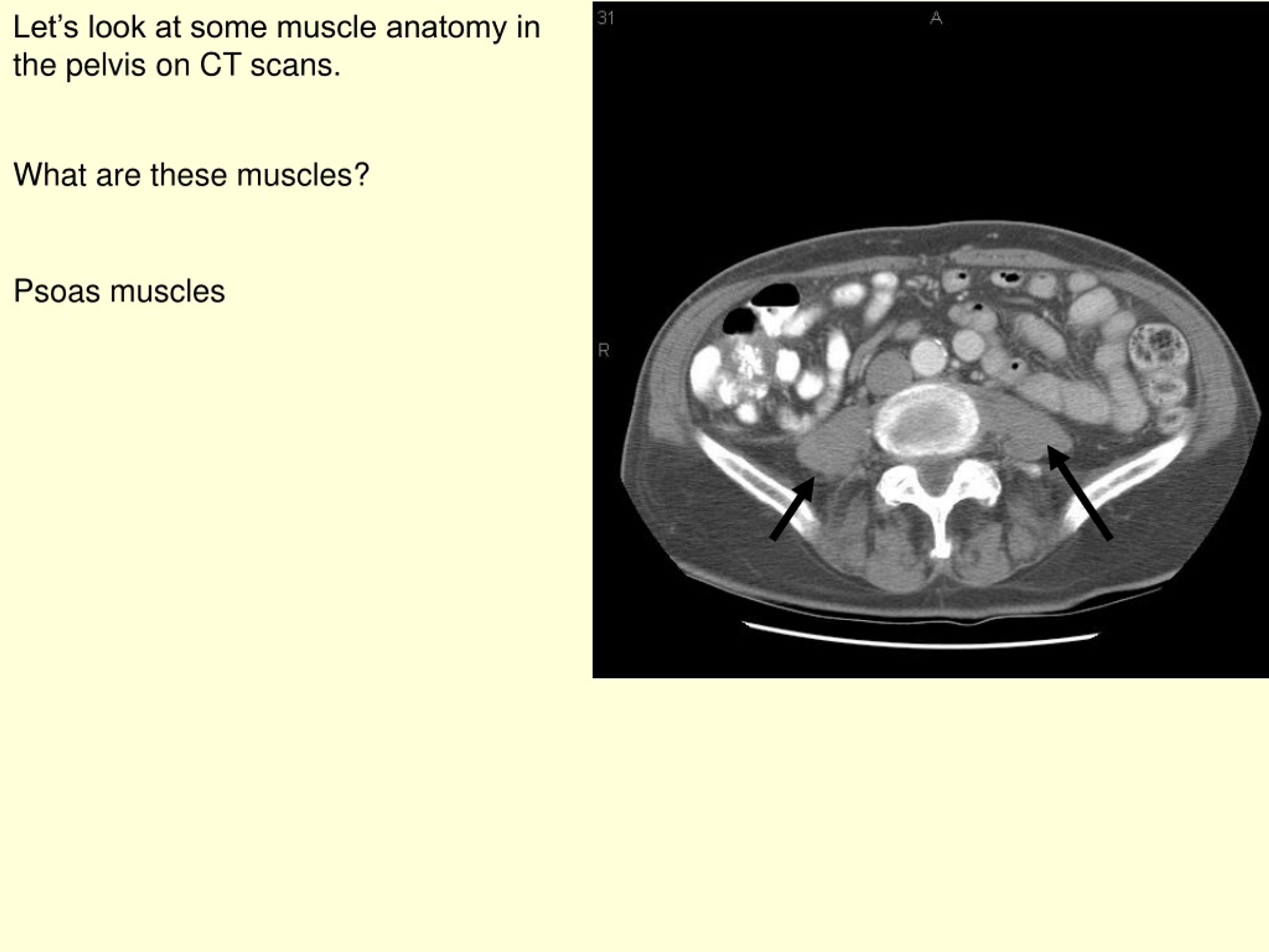
Ppt Let S Look At Some Muscle Anatomy In The Pelvis On Ct Scans Powerpoint Presentation Id 9114577 from image4.slideserve.com Renal pelvis or ureter cancer. Axial section through male bladder. Pelvic floor muscles that are located wholly within the pelvis. Intravenous contrast has been given. There are many muscles that form the pelvic floor, including puborectalis, pubococcygeus, iliococcygeus and coccygeus. Postnatally, the human upright posture has also placed highly species specific physical demands on this structure. • to assess equivocal imaging findings • staging of hepatic neoplasms • metastatic workup of primary malignancies • diagnosis of abdominal masses • assessment of biliary problems • diagnosis of vascular lesions. This mri male pelvis axial cross sectional anatomy tool is absolutely free to use.
Pelvic floor muscles that are located wholly within the pelvis.
Pelvic anatomy mri variant anatomy pelvic viscera. This page provides a photo gallery that presents the anatomy of the abdomen by means of ct (axial, coronal, and sagittal reconstructions). Intravenous contrast has been given. Use the mouse scroll wheel to move the images up and down alternatively use the tiny arrows (>>) on both side of the image to move the images. It affects the entire lower limb and the movement of the hip and the lumbar area. The full bladder displaces small bowel loops superiorly. They support the pelvic organs especially during increases in intra abdominal pressure and also aid in urinary and faecal. We study anatomy at the practical anatomy class we study the human body. The muscles are connected with the bones. Although ultrasound is frequently indicated for the primary. The pelvis is a basin shaped bony structure formed by the combination of two pelvic bones (hip bones or innominate bones) and the sacrum. Anatomical drawing of the female pelvis. Innervation of the female levator ani muscles.
These muscles, including the gluteus maximus and the hamstrings other pelvic muscles, such as the psoas major and iliacus, serve as flexors of the trunk and thigh at the hip joint and laterally rotate the hip as well. The gastrocnemius muscle is a complex muscle that is fundamental for walking and posture. The video covers the most. It attaches to the walls of the lesser pelvis, separating the pelvic cavity from the perineum inferiorly (region which includes the in this article, we shall look at the anatomy of the muscles that make up the inferior lining of the cavity; Learn about anatomy muscles pelvis with free interactive flashcards.
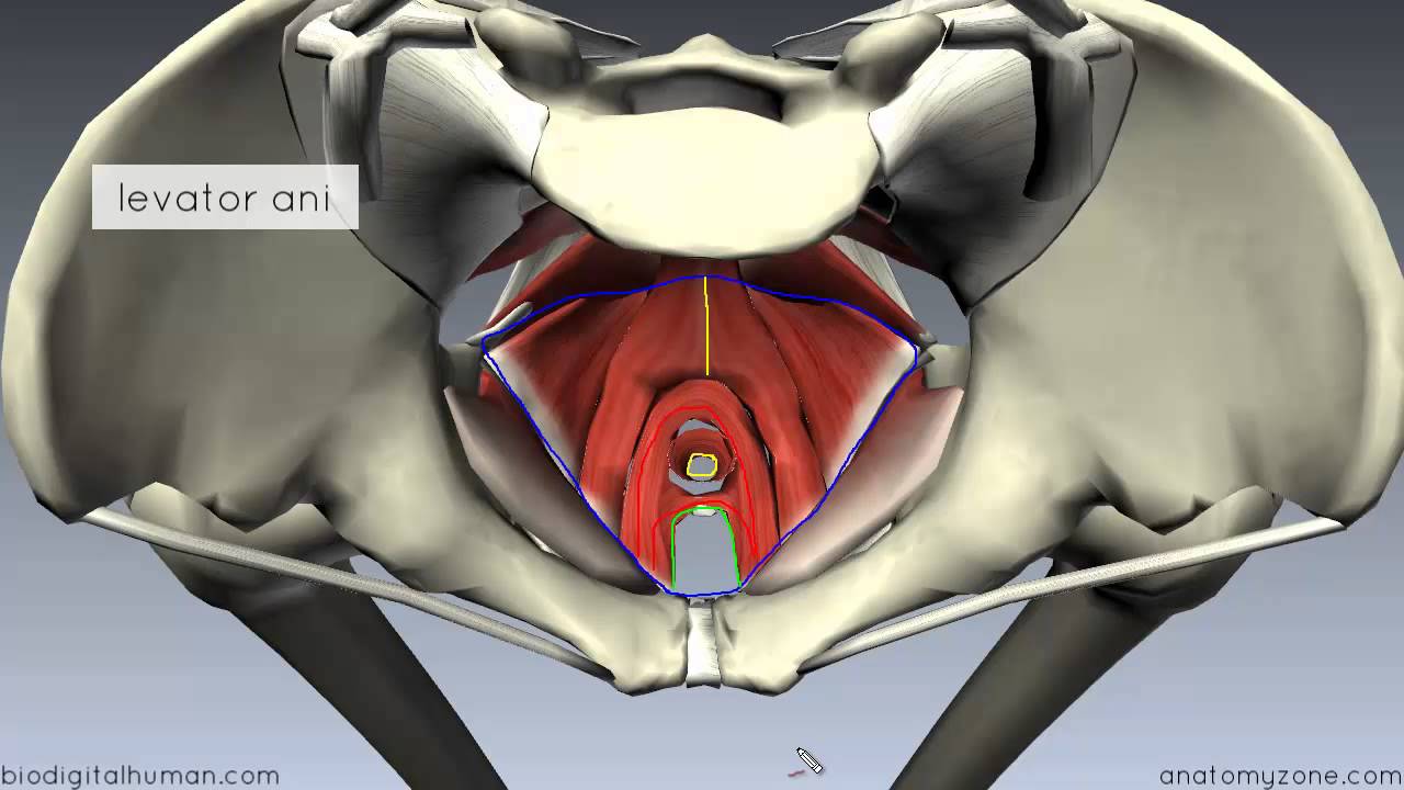
Pelvic Floor Part 1 The Pelvic Diaphragm 3d Anatomy Tutorial Youtube from i.ytimg.com This page provides a photo gallery that presents the anatomy of the abdomen by means of ct (axial, coronal, and sagittal reconstructions). The pelvis is a basin shaped bony structure formed by the combination of two pelvic bones (hip bones or innominate bones) and the sacrum. Axial section through male bladder. 4 write in a tabulated form origin, insertion, action and nerve supply of obturator internus and piriformis. Anatomical drawing of the female pelvis. Learn about anatomy muscles pelvis with free interactive flashcards. Ct anatomy of the pelvis. (2) the levator ani and the coccygeus, which together form the pelvic diaphragm and are.
Learn about anatomy muscles pelvis with free interactive flashcards.
Intravenous contrast has been given. Although ultrasound is frequently indicated for the primary. The muscles are connected with the bones. The muscles of the pelvis form its floor. This article reviews the anatomical and functional information of the gastrocnemius muscle, its embryological derivation. Muscles of the pelvis that cross the lumbosacral joint to attach onto the trunk were described in the previous blog post note: Rib thorax lumbar pelvis sacrum coccyx femur fibula tibia. Attached to the pelvis are muscles of the buttocks, the lower back, and the thighs. (1) the obturator internus and the piriformis, which are muscles of the lower extremity, and will be described with these (pages 476 and 477); Functional anatomy of the male pelvicfloor explore the important aspects of the structures and functions of the male pelvic. Functional anatomy of the male pelvic floor online course: Pelvic floor muscles that are located wholly within the pelvis. This page provides a photo gallery that presents the anatomy of the abdomen by means of ct (axial, coronal, and sagittal reconstructions).
Online mri & ct sectional anatomy. 3 enumerate the muscles of true pelvis. Attached to the pelvis are muscles of the buttocks, the lower back, and the thighs. Postnatally, the human upright posture has also placed highly species specific physical demands on this structure. The muscles within the pelvis may be divided into two groups:
Mri Anatomy Of Hip Joint Free Mri Axial Hip Anatomy from mrimaster.com • to assess equivocal imaging findings • staging of hepatic neoplasms • metastatic workup of primary malignancies • diagnosis of abdominal masses • assessment of biliary problems • diagnosis of vascular lesions. The gastrocnemius muscle is a complex muscle that is fundamental for walking and posture. This mri male pelvis axial cross sectional anatomy tool is absolutely free to use. Architectural differences in the bony pelvis of women with and without pelvic floor disorders. Pelvic floor muscles that are located wholly within the pelvis. Although ultrasound is frequently indicated for the primary. This article reviews the anatomical and functional information of the gastrocnemius muscle, its embryological derivation. Pelvic anatomy mri variant anatomy pelvic viscera.
The pelvic region holds major organs under its layers of muscles.
It affects the entire lower limb and the movement of the hip and the lumbar area. (1) the obturator internus and the piriformis, which are muscles of the lower extremity, and will be described with these (pages 476 and 477); Postnatally, the human upright posture has also placed highly species specific physical demands on this structure. Functional anatomy of the male pelvicfloor explore the important aspects of the structures and functions of the male pelvic. The video covers the most. Muscles of the pelvis that cross the lumbosacral joint to attach onto the trunk were described in the previous blog post note: This mri male pelvis axial cross sectional anatomy tool is absolutely free to use. Intravenous contrast has been given. N patient preparation n patient position n scanogram. If you want to learn how to read ct scans of the abdomen and pelvis proficiently, this video is an excellent starting point. We study anatomy at the practical anatomy class we study the human body. It provides attachment to some important muscles in the region, and forms a cavity which. Pelvic anatomy mri variant anatomy pelvic viscera.
Intravenous contrast has been given anatomy muscles pelvis. Renal pelvis or ureter cancer.


0 Komentar